


Many protozoa live in the human body. A large number of them are pathogens. Our story is about ten of them, the most. This review is based on historical and recent publications.
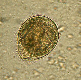
Largest. BalantidiumBalantidium coli
The largest protozoan is the human parasite, and the only ciliate in the company. Its dimensions vary from 30 to 150 microns long and from 25 to 120 microns wide. For comparison: the length of plasmodium malaria at its greatest level is about 15 microns, and several times less than the balantidium of intestinal cells, among which live infusoria. An elephant in a Chinese shop.
Distributedwherever there are pigs - their main carrier. It usually lives in the submucosa of the large intestine, although in humans it also occurs in the pulmonary epithelium. It eats bacteriaB. coli, food particles, fragments of host epithelium. In animals, the infection is not symptomatic. People can experience severe diarrhea with bloody and slimy discharge (balantidiasis), sometimes boils form on the large intestinal wall. It rarely dies from balantidiasis, but causes chronic fatigue.
People become infected through dirty water or food containing cysts. The rate of infection in humans does not exceed 1%, while pigs can be infected worldwide.
Treatedwith antibiotics, there have been no reports of drug resistance to the disease.
Discoveredby Swedish scientist Malstem in 1857. Today, balantidiasis is associated with tropical and subtropical regions, poverty and poor hygiene.
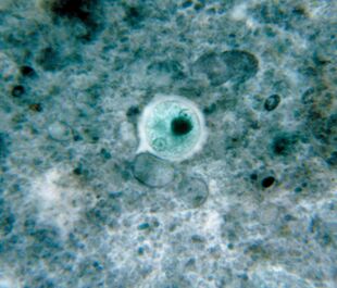
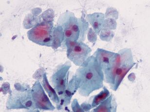
The first. Amoeba lisanEntamoeba gingivalis
Amuba is the first parasite found in humans. The description of amuba was published in 1849 in the oldest scientific journal. Amoeba is found in dental plaques, hence the name from the Latin gingivae - gum.
Alivein the mouth of almost everyone who suffers from toothache or sore gums, occupies gum pockets and plaques. It feeds on epithelial cells, leukocytes, microbes, and in the case of erythrocytes. It is rare in people with healthy mouths.
These small protozoa, measuring 10–35 µm, do not escape into the environment and do not form cysts; it is spread to other hosts through kissing, through dirty dishes or contaminated food. E. gingivalisis considered exclusively a human parasite, but is sometimes found in captive cats, dogs, horses and monkeys.
In the early twentieth century,E. gingivaliswas described as a causative agent of periodontal disease, as it is often present in inflamed dental cells. However, its pathogenicity has not been proven.
Drugsthat affect this amuba are unknown.
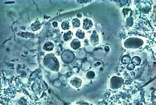
The most widespread. Dysentery amoebaEntamoeba histolytica
These intestinal parasites with blood penetrate the tissues of the liver, lungs, kidneys, brain, heart, spleen, genitals. He eats what he will get: food particles, bacteria, erythrocytes, leukocytes and epithelial cells.
Distributedeverywhere, especially in the tropics. Usually, people are infected by swallowing cysts.
In temperate countries, amuba tends to live in the intestinal lumen and the infection is not symptomatic. In the tropics and subtropics, pathological processes often begin:E. histolyticainvades the wall. The reasons for the transition to the pathogen form are still unclear, but some molecular mechanisms of what happens have been explained. Thus, it is clear that amuba releases lysing, penetrates mucus and kills cells. Apparently, amuba can destroy host cells in two ways: by triggering apoptosis in them or simply by chewing on pieces. The first method is considered the only one for a long time. By the way, the cellular suicide mechanism with record speed - in minutes - has not been identified. The second method is explained recently, the author refers to it as trogocytosis from the Greek "three" - to gnaw. It should be noted that the amuba that bites the cell leaves its prey as soon as it dies. Others can completely kill cell phagocytosis. It is assumed that biting and eating cells differ in gene expression patterns.
Now the ability of amuba to permeate into the bloodstream, liver and other organs is associated with trohocytosis.
Amoebiasis is a deadly disease, about 100 thousand people die from infection withE. histolyticaevery year.
Amuba dysentery has non-pathogenic twins,E. dispar, so microscopy is not enough to diagnose the disease.
To healmust be destroyed for mobileE. histolyticaand cysts.
ExplainedE. histolyticaand determined its pathogenic properties in 1875 in a patient with diarrhea. The Latin name for amuba was given in 1903 by German zoologist Fritz Schaudin.Histolyticameans damaged tissue. In 1906, the scientist died of an amuba intestinal abscess.
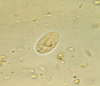
The most common. Lamblia intestineGiardia lamblia (G. intestinalis)
Giardia, the most common intestinal parasite, is found everywhere. 3-7% of people in developed countries and 20-30% in developing countries are infected. That is about 300 million people.
Living parasitesin the host duodenum and bile duct, where they float, work with the flagella, then they attach to the epithelium with the help of a sticky disk located at the bottom of the cell. For 1 cm2, the epithelium adheres to one million lamblia. They damage the villi, which interferes with the absorption of nutrients, causing inflammation of the mucosa and diarrhea. If the disease affects the bile ducts, it is accompanied by jaundice.
Giardiasis is a disease of the hands, water and dirty food. The life cycle of protozoa is simple: in the intestine there is an active form, and at the exit with a stool mass, the cyst is stable. To be infected, it is enough to swallow a dozen cysts, which in the intestine will return to active form.
The main secretis the presence of lamblia in surface protein variability. The human body fights lamblia with antibodies and, basically, is able to develop immunity. But people who live in the same area and drink the same water are infected repeatedly by their own parasitic offspring. Why? Because during the transition from the active phase to the cyst and vice versa, lamblia convert to proteins in which antibodies are produced - variant-specific surface proteins. There are about 190 variants of this protein in the genome, but only one is found on the surface of individual parasites; the rest of the translation is interrupted by the mechanism of RNA interference. And that change happened about ten generations at a time.
It is treatedwith antiprotozoal agents with antibacterial activity. The disease disappears within a week, but if the bile ducts are infected, relapse may occur over many years. Cysts are fought by ionizing water.
FoundGiardia lambliain 1859 by Czech scientist Vilém Lambl. Since then, the most modest have changed some names and are now accepted in honor of the French inventor and parasite Alfred Giar, who does not describe lamblia.
And the first sketch of Giardia was made by Anthony van Leeuwenhoek, who found it in his disappointed chair. In 1681.
By the way, Giardia is also very ancient in evolution, it comes almost directly from the ancestors of all eukaryotes.
The most intimate. Trichomonas vaginalisTrichomonas vaginalis.
The simplest, sexually disseminated. It lives in the vagina, and in men - in the urethra, epididymis and prostate gland, it is sexually transmitted or through wet wipes. Babies can be infected through the birth canal.T. vaginalishas 4 flagella at the anterior end and a relatively short corrugated membrane; if necessary, it releases a pseudopod. The maximum size of Trichomonas is 32 x 12 microns.
Trichomonas is morewidespreadthan the causative agents of chlamydia, gonorrhea and syphilis. It affects about 10% of women, and possibly more, and 1% of men. The last figure is unreliable because it is more difficult to detect parasites in men.
T. vaginaliseats microorganisms, including lactic acid bacteria of the vaginal microflora, which maintain an acidic environment, and thus create an optimal pH for itself above 4. 9.
Trichomonas damages mucosal cells, causing inflammation. About 15% of infected women complain of symptoms.
It is treatedwith antibacterial drugs. As a preventative measure, regular washing with dilute vinegar is recommended.
Explainedin 1836 by French bacteriologist Alfred Donne. Scientists do not understand that there are pathogenic parasites in front of him, but he determines the simplest size, appearance and type of movement.
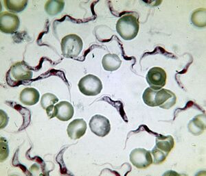
The most deadly. The causative agent of sleep disordersTrypanosoma brucei
The causative agent of African sleep disorders is the most deadly protozoa. An infected person dies without treatment. Trypanosoma is a flagellate length of 15-40 µm. Two known subspecies are outwardly indistinguishable. Diseases caused byT. brucei gambiense, lasts 2-4 years.T. brucei rhodesienseis a more dangerous and temporary pathogen from which they die after a few months or weeks.
Distributedin Africa, between the 15th parallel of the Southern and Northern Hemispheres, in the natural range of carriers - blood-sucking insects of the genusGlossina(tsetse fly). Of the 31 species of flies, 11 are dangerous to humans. Sleep disorders affect the population of 37 countries in the southern Sahara with a distance of 9 million km2. Up to 20, 000 people get sick every year. There are now about 500 thousand patients, 60 million living at risk.
From the intestines of fliesT. bruceienters the human bloodstream, from there it enters the cerebrospinal fluid and affects the nervous system. The disease begins with fever and inflammation of the lymph nodes, followed by lethargy, drowsiness, muscle paralysis, fatigue and irreversible coma.
The death of a parasite is associated with its ability to cross the blood-brain barrier. The molecular mechanism is not fully understood, but it is known that when entering the brain, parasites secrete cysteine proteases and even use some host proteins. In the central nervous system, the trypanosome is sheltered from immune factors.
The first description of sleep disorders in the upper Niger was made by the Arab scholar Ibn Khaldun (1332-1406). By the early 19th century, Europeans already knew the early signs of the disease - swelling of the lymph nodes in the back of the neck (Winterbottom symptoms), and slave traders paid special attention to it.
FoundT. bruceiScottish microbiologist David Bruce, after his name, was named, and in 1903 he first established a connection between the trypanosome, tsetse fly and sleep.
Treatmentdepends on the stage of the disease, medications cause severe side effects. Parasites have high antigenic variability, so it is impossible to make a vaccine.
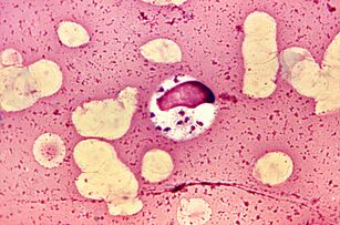
The most luxurious. LeishmaniaLeishmania donovani
Leishmania has earned the title of the most luxurious parasite, as it lives and reproduces in macrophages - cells designed to destroy parasites.L. donovaniis the most dangerous of them. It causes visceral leishmaniasis, dumdum fever, or azar, from which almost all patients die without treatment. But survivors acquire long-term immunity.
There are three subspecies of parasites.L. donovani infantum(Mediterranean and Central Asia) mainly affects children, dogs are often the reservoir.L. donovani donovani(India and Bangladesh) is dangerous for adults and the elderly, has no natural reservoir. AmericansL. donovani chagasi(Central and South America) can live in dog blood.
L. donovani- flagellate no more than 6 microns in length. People become infected after being bitten by mosquitoes of the genusPhlebotomus, sometimes through sexual intercourse, infants - through the birth canal. Once in the blood,L. donovanipermeates into macrophages, which carry parasites through internal organs. Breeding in macrophages, parasites destroy it. The mechanism of molecular survival in macrophages is quite complex.
Symptoms of the disease- fever, enlarged liver and spleen, anemia and leukopenia, which contribute to secondary bacterial infections. Every year 500 thousand people fall ill with visual leishmaniasis and about 40 thousand die.
Treatmentweight - intravenous antimony and blood travel.
Combined taxonomyL. donovaniwas determined in 1903 by renowned malaria researcher and Nobel laureate Ronald Ross. This is thanks to his generic name to William Leishman, and a specific name for Charles Donovan, who in 1903 found the same protozoa cells freely in the spleen of patients who died of azar, one in London, the other in Madras.
The most difficult life cycle.Babesia spp.
Babesias, in addition to elevated asexual reproduction in mammalian erythrocytes and sexual mites in the intestine genusIxodes, complicates its development through transovarial transmission. From the intestines of female mites, sporozoite protozoa penetrate the ovaries and infect the embryo. When mite larvae hatch, babesia enter their salivary glands and, with the first bite, enter vertebrate blood.
DistributedBabesia in America, Europe and Asia. Their natural reservoirs are rats, dogs and cattle. A person is infected with several types: B. microti, B. divergens, B. duncanidanB. venatorum.
Symptoms of babesiosis are similar to malaria - recurrent fever, hemolytic anemia, enlarged spleen and liver. A large number of people recover spontaneously, but babesiosis is fatal to patients with a weakened immune system.
Methods of treatmentare still in development, while antibiotics are prescribed and, in severe cases, blood transfusions.
Babesia was described by Romanian microbiologist Victor Babes (1888), who found it in sick cows and sheep. He decided that he was dealing with a pathogenic bacterium he namedHaematococcus bovis. Babesia has long been considered an animal pathogen until it was discovered in 1957 on a Yugoslavian shepherd who died of B. divergens infection.
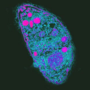
The most influential. The causative agent of toxoplasmosisToxoplasma gondii
T. gondiiis the most powerful parasite as it controls the behavior of intermediate hosts.
Distributedeverywhere, distributed unevenly. In France, for example, 84% of the population is infected, in the United Kingdom - 22%.
Toxoplasma life cycle consists of two stages: asexual occurs in the body of any hot blood, sexual reproduction is possible only in the intestinal epithelial cells of cats. ToT. gondiican complete the development, the cat must eat an infected rat. By increasing the likelihood of this occurrence,T. gondiisuppresses the rat's natural fear of cat urine odor and makes it attractive by targeting a group of neurons in the amygdala. How he did it is unknown. One of the proper mechanisms of action is the local immune response to infection. It alters cytokine levels, which in turn increase the levels of neuromodulators such as dopamine. Toxoplasm also affects human behavior, which is manifested even at the population level. So, in countries with high levels of toxoplasmosis, neuroticism and the desire to avoid uncertainty, new situations are more common. It is possible that infection withT. gondiican cause cultural changes.
Infectionsin humans are often asymptomatic, but with weakened immunity, they damage the cells of the liver, lungs, brain, retina, causing acute or chronic toxoplasmosis. Infection depends on the severity of the strain, the condition of the host's immune system and its age - older people are less susceptible toT. gondii.
Treattoxoplasmosis with antiprotozoal drugs.
Explainedin 1908 on desert mice. This honor belongs to the staff of the Pasteur Institute in Tunisia Charles Nicolas and Luis Manso.
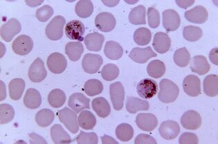
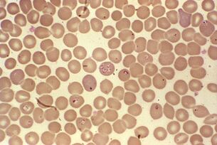
Most pathogens. Plasmodium malariaPlasmodium spp.
Plasmodium malaria is the most pathogenic parasite in humans. The number of patients with malaria can reach 300-500 million, and the mortality rate during the epidemic - 2 million. The disease still claimed three times more lives than armed conflict.
Five species of Plasmodium cause malaria in humans:Plasmodium vivax, P. falciparum, P. malariae, P. ovaleandP. knowlesi, which also affect monkeys.
Distributedin the range of vectors - mosquitoesAnopheles, which require a temperature of 16-34 ° C and a relative humidity of more than 60%.
A comparison of the most violent plasmodia genome,P. falciparum, with the gorilla plasmodia shows that humans are infected by their ancestors from these monkeys. The emergence of this form of Plasmodium is associated with the emergence of agriculture in Africa, which led to an increase in population density and the development of irrigation systems.
Sexual reproduction of plasmodia occurs in the intestines of mosquitoes, and in the human body it is an intracellular parasite that lives and multiplies in hepatocytes and erythrocytes until cells break down. 1 ml of patient blood contains 1 - 50 thousand parasites.
The disease manifests itself as inflammation, recurrent fever and anemia, if pregnant it is dangerous for both mother and fetus. Infected erythrocytesP. falciparumclog capillaries, and in severe cases ischemia of organs and internal tissues develops.
Treatmentrequires a combination of several drugs and depends on the specific pathogen. Plasmodia becomes drug resistant.